Publication date: Available online 10 June 2016
Source:Gait & Posture
Author(s): Maxime T. Robert, Laurent Ballaz, Martin Lemay
IntroductionIn the next few years, several head-mounted displays (HMD) will be publicly released making virtual reality more accessible. HMD are expected to be widely popular at home for gaming but also in clinical settings, notably for training and rehabilitation. HMD can be used in both seated and standing positions; however, presently, the impact of HMD on balance remains largely unknown. It is therefore crucial to examine the impact of viewing a virtual environment through a HMD on standing balance.ObjectivesTo compare static and dynamic balance in a virtual environment perceived through a HMD and the physical environment. The visual representation of the virtual environment was based on filmed image of the physical environment and was therefore highly similar.DesignThis is an observational study in healthy adults.ResultsNo significant difference was observed between the two environments for static balance. However, dynamic balance was more perturbed in the virtual environment when compared to that of the physical environment.ConclusionsHMD should be used with caution because of its detrimental impact on dynamic balance. Sensorimotor conflict possibly explains the impact of HMD on balance.
from #Audiology via ola Kala on Inoreader http://ift.tt/1Og9Yty
via IFTTT
Παρασκευή 10 Ιουνίου 2016
In vivo kinematic study of the tarsal joints complex based on fluoroscopic 3D-2D registration technique
Publication date: Available online 10 June 2016
Source:Gait & Posture
Author(s): M.D. Chen Wang, Xiang Geng, Shaobai Wang, M.D. Xin Ma, M.D. Xu Wang, M.D. Jiazhang Huang, M.D. Chao Zhang, M.S. Li Chen, Junsheng Yang, Kan Wang
The tarsal bones articulate with each other and demonstrate complicated kinematic characteristics. The in vivo motions of these tarsal joints during normal gait are still unclear. Seven healthy subjects were recruited and fourteen feet in total were tested in current study. Three dimensional models of the tarsal bones were first created using CT scanning. Corresponding local 3D coordinate systems of each tarsal bone was subsequently established for 6DOF motion decompositions. The fluoroscopy system captured the lateral fluoroscopic images of the targeted tarsal region whilst the subject was walking. Seven key pose images during the stance phase were selected and 3D to 2D bone model registrations were performed on each image to determine joint positions. The 6DOF motions of each tarsal joint during gait were then obtained by connecting these positions together. The TNJ (talo-navicular joint) exhibited the largest ROMs (range of motion) on all rotational directions with 7.39±2.75°of dorsi/plantarflexion, 21.12±4.68°of inversion/eversion, and 16.11±4.44°of internal/external rotation. From heel strike to midstance, the TNJ, STJ (subtalar joint), and CCJ (calcaneao-cuboid joint) were associated with 5.97°, 5.04°, and 3.93°of dorsiflexion; 15.46°, 8.21°, and 5.82°of eversion; and 9.75°, 7.6°, and 4.99°of external rotation, respectively. Likewise, from midstance to heel off, the TNJ, STJ, and CCJ were associated with 6.39, 6.19°, and 4.47°of plantarflexion; 18.57°, 11.86°, and 6.32°of inversion and 13.95°, 9.66°, and 7.58°of internal rotation, respectively. In conclusion, among the tarsal joints, the TNJ exhibited the greatest rotational mobility. Synchronous and homodromous rotational motions were detected for TNJ, STJ, and CCJ during the stance phase.
from #Audiology via ola Kala on Inoreader http://ift.tt/1tgiRtm
via IFTTT
The effect of viewing a virtual environment through a head-mounted display on balance
Publication date: Available online 10 June 2016
Source:Gait & Posture
Author(s): Maxime T. Robert, Laurent Ballaz, Martin Lemay
IntroductionIn the next few years, several head-mounted displays (HMD) will be publicly released making virtual reality more accessible. HMD are expected to be widely popular at home for gaming but also in clinical settings, notably for training and rehabilitation. HMD can be used in both seated and standing positions; however, presently, the impact of HMD on balance remains largely unknown. It is therefore crucial to examine the impact of viewing a virtual environment through a HMD on standing balance.ObjectivesTo compare static and dynamic balance in a virtual environment perceived through a HMD and the physical environment. The visual representation of the virtual environment was based on filmed image of the physical environment and was therefore highly similar.DesignThis is an observational study in healthy adults.ResultsNo significant difference was observed between the two environments for static balance. However, dynamic balance was more perturbed in the virtual environment when compared to that of the physical environment.ConclusionsHMD should be used with caution because of its detrimental impact on dynamic balance. Sensorimotor conflict possibly explains the impact of HMD on balance.
from #Audiology via xlomafota13 on Inoreader http://ift.tt/1Og9Yty
via IFTTT
In vivo kinematic study of the tarsal joints complex based on fluoroscopic 3D-2D registration technique
Publication date: Available online 10 June 2016
Source:Gait & Posture
Author(s): M.D. Chen Wang, Xiang Geng, Shaobai Wang, M.D. Xin Ma, M.D. Xu Wang, M.D. Jiazhang Huang, M.D. Chao Zhang, M.S. Li Chen, Junsheng Yang, Kan Wang
The tarsal bones articulate with each other and demonstrate complicated kinematic characteristics. The in vivo motions of these tarsal joints during normal gait are still unclear. Seven healthy subjects were recruited and fourteen feet in total were tested in current study. Three dimensional models of the tarsal bones were first created using CT scanning. Corresponding local 3D coordinate systems of each tarsal bone was subsequently established for 6DOF motion decompositions. The fluoroscopy system captured the lateral fluoroscopic images of the targeted tarsal region whilst the subject was walking. Seven key pose images during the stance phase were selected and 3D to 2D bone model registrations were performed on each image to determine joint positions. The 6DOF motions of each tarsal joint during gait were then obtained by connecting these positions together. The TNJ (talo-navicular joint) exhibited the largest ROMs (range of motion) on all rotational directions with 7.39±2.75°of dorsi/plantarflexion, 21.12±4.68°of inversion/eversion, and 16.11±4.44°of internal/external rotation. From heel strike to midstance, the TNJ, STJ (subtalar joint), and CCJ (calcaneao-cuboid joint) were associated with 5.97°, 5.04°, and 3.93°of dorsiflexion; 15.46°, 8.21°, and 5.82°of eversion; and 9.75°, 7.6°, and 4.99°of external rotation, respectively. Likewise, from midstance to heel off, the TNJ, STJ, and CCJ were associated with 6.39, 6.19°, and 4.47°of plantarflexion; 18.57°, 11.86°, and 6.32°of inversion and 13.95°, 9.66°, and 7.58°of internal rotation, respectively. In conclusion, among the tarsal joints, the TNJ exhibited the greatest rotational mobility. Synchronous and homodromous rotational motions were detected for TNJ, STJ, and CCJ during the stance phase.
from #Audiology via xlomafota13 on Inoreader http://ift.tt/1tgiRtm
via IFTTT
Genetic, Clinical, and Pathologic Backgrounds of Patients with Autosomal Dominant Alport Syndrome.
Genetic, Clinical, and Pathologic Backgrounds of Patients with Autosomal Dominant Alport Syndrome.
Clin J Am Soc Nephrol. 2016 Jun 8;
Authors: Kamiyoshi N, Nozu K, Fu XJ, Morisada N, Nozu Y, Ye MJ, Imafuku A, Miura K, Yamamura T, Minamikawa S, Shono A, Ninchoji T, Morioka I, Nakanishi K, Yoshikawa N, Kaito H, Iijima K
Abstract
BACKGROUND AND OBJECTIVES: Alport syndrome comprises a group of inherited heterogeneous disorders involving CKD, hearing loss, and ocular abnormalities. Autosomal dominant Alport syndrome caused by heterozygous mutations in collagen 4A3 and/or collagen 4A4 accounts for <5% of patients. However, the clinical, genetic, and pathologic backgrounds of patients with autosomal dominant Alport syndrome remain unclear.
DESIGN, SETTING, PARTICIPANTS, & MEASUREMENTS: We conducted a retrospective analysis of 25 patients with genetically proven autosomal dominant Alport syndrome and their family members (a total of 72 patients) from 16 unrelated families. Patients with suspected Alport syndrome after pathologic examination who were referred from anywhere in Japan for genetic analysis from 2006 to 2015 were included in this study. Clinical, laboratory, and pathologic data were collected from medical records at the point of registration for genetic diagnosis. Genetic analysis was performed by targeted resequencing of 27 podocyte-related genes, including Alport-related collagen genes, to make a diagnosis of autosomal dominant Alport syndrome and identify modifier genes or double mutations. Clinical data were obtained from medical records.
RESULTS: The median renal survival time was 70 years, and the median age at first detection of proteinuria was 17 years old. There was one patient with hearing loss and one patient with ocular lesion. Among 16 patients who underwent kidney biopsy, three showed FSGS, and seven showed thinning without lamellation of the glomerular basement membrane. Five of 13 detected mutations were reported to be causative mutations for autosomal recessive Alport syndrome in previous studies. Two families possessed double mutations in both collagen 4A3 and collagen 4A4, but no modifier genes were detected among the other podocyte-related genes.
CONCLUSIONS: The renal phenotype of autosomal dominant Alport syndrome was much milder than that of autosomal recessive Alport syndrome or X-linked Alport syndrome in men. It may, thus, be difficult to make an accurate diagnosis of autosomal dominant Alport syndrome on the basis of clinical or pathologic findings. No modifier genes were identified among the known podocyte-related genes.
PMID: 27281700 [PubMed - as supplied by publisher]
from #Audiology via xlomafota13 on Inoreader http://ift.tt/25RcMSr
via IFTTT
RIN2 syndrome: Expanding the clinical phenotype.
RIN2 syndrome: Expanding the clinical phenotype.
Am J Med Genet A. 2016 Jun 8;
Authors: Rosato S, Syx D, Ivanovski I, Pollazzon M, Santodirocco D, De Marco L, Beltrami M, Callewaert B, Garavelli L, Malfait F
Abstract
Biallelic defects in the RIN2 gene, encoding the Ras and Rab interactor 2 protein, are associated with a rare autosomal recessive connective tissue disorder, with only nine patients from four independent families reported to date. The condition was initially termed MACS syndrome (macrocephaly, alopecia, cutis laxa, and scoliosis), based on the clinical features of the first identified family; however, with the expansion of the clinical phenotype in additional families, it was subsequently coined RIN2 syndrome. Hallmark features of this condition include dysmorphic facial features with striking, progressive facial coarsening, sparse hair, normal to enlarged occipitofrontal circumference, soft redundant and/or hyperextensible skin, and scoliosis. Patients with RIN2 syndrome present phenotypic overlap with other conditions, including EDS (especially the dermatosparaxis and kyphoscoliosis subtypes). Here, we describe a 10th patient, the first patient of Caucasian origin and the oldest reported patient so far, who harbors the previously identified homozygous RIN2 mutation c.1878dupC (p. (Ile627Hisfs*7)). Besides the hallmark features, this patient also presents problems not previously associated with RIN2 syndrome, including cervical vertebral fusion, mild hearing loss, and colonic fibrosis. We provide an overview of the clinical findings in all reported patients with RIN2 mutations and summarize some of the possible pathogenic mechanisms that may underlie this condition. © 2016 Wiley Periodicals, Inc.
PMID: 27277385 [PubMed - as supplied by publisher]
from #Audiology via xlomafota13 on Inoreader http://ift.tt/1XKOUii
via IFTTT
The effect of viewing a virtual environment through a head-mounted display on balance
Publication date: Available online 10 June 2016
Source:Gait & Posture
Author(s): Maxime T. Robert, Laurent Ballaz, Martin Lemay
IntroductionIn the next few years, several head-mounted displays (HMD) will be publicly released making virtual reality more accessible. HMD are expected to be widely popular at home for gaming but also in clinical settings, notably for training and rehabilitation. HMD can be used in both seated and standing positions; however, presently, the impact of HMD on balance remains largely unknown. It is therefore crucial to examine the impact of viewing a virtual environment through a HMD on standing balance.ObjectivesTo compare static and dynamic balance in a virtual environment perceived through a HMD and the physical environment. The visual representation of the virtual environment was based on filmed image of the physical environment and was therefore highly similar.DesignThis is an observational study in healthy adults.ResultsNo significant difference was observed between the two environments for static balance. However, dynamic balance was more perturbed in the virtual environment when compared to that of the physical environment.ConclusionsHMD should be used with caution because of its detrimental impact on dynamic balance. Sensorimotor conflict possibly explains the impact of HMD on balance.
from #Audiology via ola Kala on Inoreader http://ift.tt/1Og9Yty
via IFTTT
In vivo kinematic study of the tarsal joints complex based on fluoroscopic 3D-2D registration technique
Publication date: Available online 10 June 2016
Source:Gait & Posture
Author(s): M.D. Chen Wang, Xiang Geng, Shaobai Wang, M.D. Xin Ma, M.D. Xu Wang, M.D. Jiazhang Huang, M.D. Chao Zhang, M.S. Li Chen, Junsheng Yang, Kan Wang
The tarsal bones articulate with each other and demonstrate complicated kinematic characteristics. The in vivo motions of these tarsal joints during normal gait are still unclear. Seven healthy subjects were recruited and fourteen feet in total were tested in current study. Three dimensional models of the tarsal bones were first created using CT scanning. Corresponding local 3D coordinate systems of each tarsal bone was subsequently established for 6DOF motion decompositions. The fluoroscopy system captured the lateral fluoroscopic images of the targeted tarsal region whilst the subject was walking. Seven key pose images during the stance phase were selected and 3D to 2D bone model registrations were performed on each image to determine joint positions. The 6DOF motions of each tarsal joint during gait were then obtained by connecting these positions together. The TNJ (talo-navicular joint) exhibited the largest ROMs (range of motion) on all rotational directions with 7.39±2.75°of dorsi/plantarflexion, 21.12±4.68°of inversion/eversion, and 16.11±4.44°of internal/external rotation. From heel strike to midstance, the TNJ, STJ (subtalar joint), and CCJ (calcaneao-cuboid joint) were associated with 5.97°, 5.04°, and 3.93°of dorsiflexion; 15.46°, 8.21°, and 5.82°of eversion; and 9.75°, 7.6°, and 4.99°of external rotation, respectively. Likewise, from midstance to heel off, the TNJ, STJ, and CCJ were associated with 6.39, 6.19°, and 4.47°of plantarflexion; 18.57°, 11.86°, and 6.32°of inversion and 13.95°, 9.66°, and 7.58°of internal rotation, respectively. In conclusion, among the tarsal joints, the TNJ exhibited the greatest rotational mobility. Synchronous and homodromous rotational motions were detected for TNJ, STJ, and CCJ during the stance phase.
from #Audiology via ola Kala on Inoreader http://ift.tt/1tgiRtm
via IFTTT
A Case of Undiagnosed Sleep Disorder with Hearing Difficulty and Dizziness.
A Case of Undiagnosed Sleep Disorder with Hearing Difficulty and Dizziness.
Iran J Otorhinolaryngol. 2016 Mar;28(85):149-52
Authors: Goto F, Arai M, Kitamura M, Otomo T, Nagai R, Minami S, Shimada T, Matsunaga T, Tsunoda K, Fujii M
Abstract
INTRODUCTION: The aim of this case report was to investigate the relationship between sleep disorders and audio vestibular symptoms.
CASE REPORT: A case of undiagnosed sleep disorder, presenting as a temporary auditory processing difficulty, is presented. The disorder was initially treated as sudden deafness with dizziness. A 23-year-old male patient complained of acute hearing disturbance despite normal results on pure tone audiometry. The patient was initially administered a steroid injection in the hospital. After treatment, his hearing symptoms improved only slightly and he reported balance difficulty with rightward spontaneous nystagmus. Vestibular rehabilitation was performed. We also suspected that his hearing symptom was due to an auditory processing difficulty. Despite steroid treatment and vestibular rehabilitation, neither of his symptoms improved. We subsequently identified the presence of insomnia. He was prescribed zolpidem 5 mg, which slightly improved his symptoms, and referred to a sleep specialist for further examination. Polysomnography was performed, which identified restless leg syndrome and sleep disturbance with delayed sleep phase syndrome. After pharmacological treatment, his sleep disturbance, hearing difficulty, and balance disorder completely resolved.
CONCLUSION: Sleep disorders may provoke reversible auditory processing difficulties. We should carefully evaluate patients for a potentially undiagnosed sleep disorder, even in patients chiefly complaining of intractable sensory dysfunction such as hearing or balance disturbance.
PMID: 27280102 [PubMed]
from #Audiology via ola Kala on Inoreader http://ift.tt/24F50sk
via IFTTT
Audiological Monitoring in Children Treated with Platinum Chemotherapy
Platinum compounds constitute the standard treatment for solid tumors in pediatric oncology. The purpose of this study is to assess the impact of platinum compounds in the development of ototoxicity in children following chemotherapy. This study included 160 patients treated with cisplatin and carboplatin for malignant solid diseases from 2007 to 2014. Their audiograms were classified according to the Boston SIOP ototoxicity scale. Twenty-five percent of the children treated with platinum compounds developed ototoxicity. The incidence of ototoxicity was correlated with the type of platinum derivative (i.e. cisplatin vs. carboplatin), coadministration of both drugs and concomitant cranial radiotherapy, but not with sex and age. Cumulative dose was correlated only with the cisplatin administration. Nine patients (8.6%) showed further progression of hearing impairment after the end of chemotherapy. The low rate of ototoxicity suggests the pivotal role of auditory monitoring in children treated with platinum compounds in order to be able to identify hearing loss at an early stage and to provide, jointly with pediatric oncologists, strategies to reduce further progression of cochlear toxicity.
Audiol Neurotol 2016;21:203-211
from #Audiology via xlomafota13 on Inoreader http://ift.tt/1tg5WHZ
via IFTTT
Audiol Neurotol 2016;21:203-211
from #Audiology via xlomafota13 on Inoreader http://ift.tt/1tg5WHZ
via IFTTT
Neural correlates of auditory scale illusion
Publication date: Available online 9 June 2016
Source:Hearing Research
Author(s): Shinya Kuriki, Ryousuke Numao, Iku Nemoto
The auditory illusory perception “scale illusion” occurs when ascending and descending musical scale tones are delivered in a dichotic manner, such that the higher or lower tone at each instant is presented alternately to the right and left ears. Resulting tone sequences have a zigzag pitch in one ear and the reversed (zagzig) pitch in the other ear. Most listeners hear illusory smooth pitch sequences of up-down and down-up streams in the two ears separated in higher and lower halves of the scale. Although many behavioral studies have been conducted, how and where in the brain the illusory percept is formed have not been elucidated. In this study, we conducted functional magnetic resonance imaging using sequential tones that induced scale illusion (ILL) and those that mimicked the percept of scale illusion (PCP), and we compared the activation responses evoked by those stimuli by region-of-interest analysis. We examined the effects of adaptation, i.e., the attenuation of response that occurs when close-frequency sounds are repeated, which might interfere with the changes in activation by the illusion process. Results of the activation difference of the two stimuli, measured at varied tempi of tone presentation, in the superior temporal auditory cortex were not explained by adaptation. Instead, excess activation of the ILL stimulus from the PCP stimulus at moderate tempi (83 and 126 bpm) was significant in the posterior auditory cortex with rightward superiority, while significant prefrontal activation was dominant at the highest tempo (245 bpm). We suggest that the area of the planum temporale posterior to the primary auditory cortex is mainly involved in the illusion formation, and that the illusion-related process is strongly dependent on the rate of tone presentation.
from #Audiology via ola Kala on Inoreader http://ift.tt/25QNZOn
via IFTTT
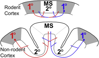
Species-dependent role of crossmodal connectivity among the primary sensory cortices
Publication date: Available online 9 June 2016
Source:Hearing Research
Author(s): M. Alex Meredith, Stephen G. Lomber
When a major sense is lost, crossmodal plasticity substitutes functional processing from the remaining, intact senses. Recent studies of deafness-induced crossmodal plasticity in different subregions of auditory cortex indicate that the phenomenon is largely based on the “unmasking” of existing inputs. However, there is not yet a consensus on the sources or effects of crossmodal inputs to primary sensory cortical areas. In the present review, a rigorous re-examination of the experimental literature indicates that connections between different primary sensory cortices consistently occur in rodents, while primary-to-primary projections are absent/inconsistent in non-rodents such as cats and monkeys. These observations suggest that crossmodal plasticity that involves primary sensory areas are likely to exhibit species-specific distinctions.
from #Audiology via ola Kala on Inoreader http://ift.tt/25QNvYA
via IFTTT
Source:Hearing Research
Author(s): M. Alex Meredith, Stephen G. Lomber
When a major sense is lost, crossmodal plasticity substitutes functional processing from the remaining, intact senses. Recent studies of deafness-induced crossmodal plasticity in different subregions of auditory cortex indicate that the phenomenon is largely based on the “unmasking” of existing inputs. However, there is not yet a consensus on the sources or effects of crossmodal inputs to primary sensory cortical areas. In the present review, a rigorous re-examination of the experimental literature indicates that connections between different primary sensory cortices consistently occur in rodents, while primary-to-primary projections are absent/inconsistent in non-rodents such as cats and monkeys. These observations suggest that crossmodal plasticity that involves primary sensory areas are likely to exhibit species-specific distinctions.
Graphical abstract
from #Audiology via ola Kala on Inoreader http://ift.tt/25QNvYA
via IFTTT
Neural correlates of auditory scale illusion
Publication date: Available online 9 June 2016
Source:Hearing Research
Author(s): Shinya Kuriki, Ryousuke Numao, Iku Nemoto
The auditory illusory perception “scale illusion” occurs when ascending and descending musical scale tones are delivered in a dichotic manner, such that the higher or lower tone at each instant is presented alternately to the right and left ears. Resulting tone sequences have a zigzag pitch in one ear and the reversed (zagzig) pitch in the other ear. Most listeners hear illusory smooth pitch sequences of up-down and down-up streams in the two ears separated in higher and lower halves of the scale. Although many behavioral studies have been conducted, how and where in the brain the illusory percept is formed have not been elucidated. In this study, we conducted functional magnetic resonance imaging using sequential tones that induced scale illusion (ILL) and those that mimicked the percept of scale illusion (PCP), and we compared the activation responses evoked by those stimuli by region-of-interest analysis. We examined the effects of adaptation, i.e., the attenuation of response that occurs when close-frequency sounds are repeated, which might interfere with the changes in activation by the illusion process. Results of the activation difference of the two stimuli, measured at varied tempi of tone presentation, in the superior temporal auditory cortex were not explained by adaptation. Instead, excess activation of the ILL stimulus from the PCP stimulus at moderate tempi (83 and 126 bpm) was significant in the posterior auditory cortex with rightward superiority, while significant prefrontal activation was dominant at the highest tempo (245 bpm). We suggest that the area of the planum temporale posterior to the primary auditory cortex is mainly involved in the illusion formation, and that the illusion-related process is strongly dependent on the rate of tone presentation.
from #Audiology via ola Kala on Inoreader http://ift.tt/25QNZOn
via IFTTT
Source:Hearing Research
Author(s): Shinya Kuriki, Ryousuke Numao, Iku Nemoto
The auditory illusory perception “scale illusion” occurs when ascending and descending musical scale tones are delivered in a dichotic manner, such that the higher or lower tone at each instant is presented alternately to the right and left ears. Resulting tone sequences have a zigzag pitch in one ear and the reversed (zagzig) pitch in the other ear. Most listeners hear illusory smooth pitch sequences of up-down and down-up streams in the two ears separated in higher and lower halves of the scale. Although many behavioral studies have been conducted, how and where in the brain the illusory percept is formed have not been elucidated. In this study, we conducted functional magnetic resonance imaging using sequential tones that induced scale illusion (ILL) and those that mimicked the percept of scale illusion (PCP), and we compared the activation responses evoked by those stimuli by region-of-interest analysis. We examined the effects of adaptation, i.e., the attenuation of response that occurs when close-frequency sounds are repeated, which might interfere with the changes in activation by the illusion process. Results of the activation difference of the two stimuli, measured at varied tempi of tone presentation, in the superior temporal auditory cortex were not explained by adaptation. Instead, excess activation of the ILL stimulus from the PCP stimulus at moderate tempi (83 and 126 bpm) was significant in the posterior auditory cortex with rightward superiority, while significant prefrontal activation was dominant at the highest tempo (245 bpm). We suggest that the area of the planum temporale posterior to the primary auditory cortex is mainly involved in the illusion formation, and that the illusion-related process is strongly dependent on the rate of tone presentation.
from #Audiology via ola Kala on Inoreader http://ift.tt/25QNZOn
via IFTTT
Species-dependent role of crossmodal connectivity among the primary sensory cortices
Publication date: Available online 9 June 2016
Source:Hearing Research
Author(s): M. Alex Meredith, Stephen G. Lomber
When a major sense is lost, crossmodal plasticity substitutes functional processing from the remaining, intact senses. Recent studies of deafness-induced crossmodal plasticity in different subregions of auditory cortex indicate that the phenomenon is largely based on the “unmasking” of existing inputs. However, there is not yet a consensus on the sources or effects of crossmodal inputs to primary sensory cortical areas. In the present review, a rigorous re-examination of the experimental literature indicates that connections between different primary sensory cortices consistently occur in rodents, while primary-to-primary projections are absent/inconsistent in non-rodents such as cats and monkeys. These observations suggest that crossmodal plasticity that involves primary sensory areas are likely to exhibit species-specific distinctions.
from #Audiology via ola Kala on Inoreader http://ift.tt/25QNvYA
via IFTTT
Source:Hearing Research
Author(s): M. Alex Meredith, Stephen G. Lomber
When a major sense is lost, crossmodal plasticity substitutes functional processing from the remaining, intact senses. Recent studies of deafness-induced crossmodal plasticity in different subregions of auditory cortex indicate that the phenomenon is largely based on the “unmasking” of existing inputs. However, there is not yet a consensus on the sources or effects of crossmodal inputs to primary sensory cortical areas. In the present review, a rigorous re-examination of the experimental literature indicates that connections between different primary sensory cortices consistently occur in rodents, while primary-to-primary projections are absent/inconsistent in non-rodents such as cats and monkeys. These observations suggest that crossmodal plasticity that involves primary sensory areas are likely to exhibit species-specific distinctions.
Graphical abstract
from #Audiology via ola Kala on Inoreader http://ift.tt/25QNvYA
via IFTTT
Neural correlates of auditory scale illusion
Source:Hearing Research
Author(s): Shinya Kuriki, Ryousuke Numao, Iku Nemoto
The auditory illusory perception “scale illusion” occurs when ascending and descending musical scale tones are delivered in a dichotic manner, such that the higher or lower tone at each instant is presented alternately to the right and left ears. Resulting tone sequences have a zigzag pitch in one ear and the reversed (zagzig) pitch in the other ear. Most listeners hear illusory smooth pitch sequences of up-down and down-up streams in the two ears separated in higher and lower halves of the scale. Although many behavioral studies have been conducted, how and where in the brain the illusory percept is formed have not been elucidated. In this study, we conducted functional magnetic resonance imaging using sequential tones that induced scale illusion (ILL) and those that mimicked the percept of scale illusion (PCP), and we compared the activation responses evoked by those stimuli by region-of-interest analysis. We examined the effects of adaptation, i.e., the attenuation of response that occurs when close-frequency sounds are repeated, which might interfere with the changes in activation by the illusion process. Results of the activation difference of the two stimuli, measured at varied tempi of tone presentation, in the superior temporal auditory cortex were not explained by adaptation. Instead, excess activation of the ILL stimulus from the PCP stimulus at moderate tempi (83 and 126 bpm) was significant in the posterior auditory cortex with rightward superiority, while significant prefrontal activation was dominant at the highest tempo (245 bpm). We suggest that the area of the planum temporale posterior to the primary auditory cortex is mainly involved in the illusion formation, and that the illusion-related process is strongly dependent on the rate of tone presentation.
from #Audiology via xlomafota13 on Inoreader http://ift.tt/25QNZOn
via IFTTT
Species-dependent role of crossmodal connectivity among the primary sensory cortices
Publication date: Available online 9 June 2016
Source:Hearing Research
Author(s): M. Alex Meredith, Stephen G. Lomber
When a major sense is lost, crossmodal plasticity substitutes functional processing from the remaining, intact senses. Recent studies of deafness-induced crossmodal plasticity in different subregions of auditory cortex indicate that the phenomenon is largely based on the “unmasking” of existing inputs. However, there is not yet a consensus on the sources or effects of crossmodal inputs to primary sensory cortical areas. In the present review, a rigorous re-examination of the experimental literature indicates that connections between different primary sensory cortices consistently occur in rodents, while primary-to-primary projections are absent/inconsistent in non-rodents such as cats and monkeys. These observations suggest that crossmodal plasticity that involves primary sensory areas are likely to exhibit species-specific distinctions.
from #Audiology via xlomafota13 on Inoreader http://ift.tt/25QNvYA
via IFTTT
Source:Hearing Research
Author(s): M. Alex Meredith, Stephen G. Lomber
When a major sense is lost, crossmodal plasticity substitutes functional processing from the remaining, intact senses. Recent studies of deafness-induced crossmodal plasticity in different subregions of auditory cortex indicate that the phenomenon is largely based on the “unmasking” of existing inputs. However, there is not yet a consensus on the sources or effects of crossmodal inputs to primary sensory cortical areas. In the present review, a rigorous re-examination of the experimental literature indicates that connections between different primary sensory cortices consistently occur in rodents, while primary-to-primary projections are absent/inconsistent in non-rodents such as cats and monkeys. These observations suggest that crossmodal plasticity that involves primary sensory areas are likely to exhibit species-specific distinctions.
Graphical abstract
from #Audiology via xlomafota13 on Inoreader http://ift.tt/25QNvYA
via IFTTT
Neural correlates of auditory scale illusion
Source:Hearing Research
Author(s): Shinya Kuriki, Ryousuke Numao, Iku Nemoto
The auditory illusory perception “scale illusion” occurs when ascending and descending musical scale tones are delivered in a dichotic manner, such that the higher or lower tone at each instant is presented alternately to the right and left ears. Resulting tone sequences have a zigzag pitch in one ear and the reversed (zagzig) pitch in the other ear. Most listeners hear illusory smooth pitch sequences of up-down and down-up streams in the two ears separated in higher and lower halves of the scale. Although many behavioral studies have been conducted, how and where in the brain the illusory percept is formed have not been elucidated. In this study, we conducted functional magnetic resonance imaging using sequential tones that induced scale illusion (ILL) and those that mimicked the percept of scale illusion (PCP), and we compared the activation responses evoked by those stimuli by region-of-interest analysis. We examined the effects of adaptation, i.e., the attenuation of response that occurs when close-frequency sounds are repeated, which might interfere with the changes in activation by the illusion process. Results of the activation difference of the two stimuli, measured at varied tempi of tone presentation, in the superior temporal auditory cortex were not explained by adaptation. Instead, excess activation of the ILL stimulus from the PCP stimulus at moderate tempi (83 and 126 bpm) was significant in the posterior auditory cortex with rightward superiority, while significant prefrontal activation was dominant at the highest tempo (245 bpm). We suggest that the area of the planum temporale posterior to the primary auditory cortex is mainly involved in the illusion formation, and that the illusion-related process is strongly dependent on the rate of tone presentation.
from #Audiology via ola Kala on Inoreader http://ift.tt/25QNZOn
via IFTTT
Species-dependent role of crossmodal connectivity among the primary sensory cortices
Publication date: Available online 9 June 2016
Source:Hearing Research
Author(s): M. Alex Meredith, Stephen G. Lomber
When a major sense is lost, crossmodal plasticity substitutes functional processing from the remaining, intact senses. Recent studies of deafness-induced crossmodal plasticity in different subregions of auditory cortex indicate that the phenomenon is largely based on the “unmasking” of existing inputs. However, there is not yet a consensus on the sources or effects of crossmodal inputs to primary sensory cortical areas. In the present review, a rigorous re-examination of the experimental literature indicates that connections between different primary sensory cortices consistently occur in rodents, while primary-to-primary projections are absent/inconsistent in non-rodents such as cats and monkeys. These observations suggest that crossmodal plasticity that involves primary sensory areas are likely to exhibit species-specific distinctions.
from #Audiology via ola Kala on Inoreader http://ift.tt/25QNvYA
via IFTTT
Source:Hearing Research
Author(s): M. Alex Meredith, Stephen G. Lomber
When a major sense is lost, crossmodal plasticity substitutes functional processing from the remaining, intact senses. Recent studies of deafness-induced crossmodal plasticity in different subregions of auditory cortex indicate that the phenomenon is largely based on the “unmasking” of existing inputs. However, there is not yet a consensus on the sources or effects of crossmodal inputs to primary sensory cortical areas. In the present review, a rigorous re-examination of the experimental literature indicates that connections between different primary sensory cortices consistently occur in rodents, while primary-to-primary projections are absent/inconsistent in non-rodents such as cats and monkeys. These observations suggest that crossmodal plasticity that involves primary sensory areas are likely to exhibit species-specific distinctions.
Graphical abstract
from #Audiology via ola Kala on Inoreader http://ift.tt/25QNvYA
via IFTTT
Neural correlates of auditory scale illusion
Source:Hearing Research
Author(s): Shinya Kuriki, Ryousuke Numao, Iku Nemoto
The auditory illusory perception “scale illusion” occurs when ascending and descending musical scale tones are delivered in a dichotic manner, such that the higher or lower tone at each instant is presented alternately to the right and left ears. Resulting tone sequences have a zigzag pitch in one ear and the reversed (zagzig) pitch in the other ear. Most listeners hear illusory smooth pitch sequences of up-down and down-up streams in the two ears separated in higher and lower halves of the scale. Although many behavioral studies have been conducted, how and where in the brain the illusory percept is formed have not been elucidated. In this study, we conducted functional magnetic resonance imaging using sequential tones that induced scale illusion (ILL) and those that mimicked the percept of scale illusion (PCP), and we compared the activation responses evoked by those stimuli by region-of-interest analysis. We examined the effects of adaptation, i.e., the attenuation of response that occurs when close-frequency sounds are repeated, which might interfere with the changes in activation by the illusion process. Results of the activation difference of the two stimuli, measured at varied tempi of tone presentation, in the superior temporal auditory cortex were not explained by adaptation. Instead, excess activation of the ILL stimulus from the PCP stimulus at moderate tempi (83 and 126 bpm) was significant in the posterior auditory cortex with rightward superiority, while significant prefrontal activation was dominant at the highest tempo (245 bpm). We suggest that the area of the planum temporale posterior to the primary auditory cortex is mainly involved in the illusion formation, and that the illusion-related process is strongly dependent on the rate of tone presentation.
from #Audiology via ola Kala on Inoreader http://ift.tt/25QNZOn
via IFTTT
Species-dependent role of crossmodal connectivity among the primary sensory cortices
Publication date: Available online 9 June 2016
Source:Hearing Research
Author(s): M. Alex Meredith, Stephen G. Lomber
When a major sense is lost, crossmodal plasticity substitutes functional processing from the remaining, intact senses. Recent studies of deafness-induced crossmodal plasticity in different subregions of auditory cortex indicate that the phenomenon is largely based on the “unmasking” of existing inputs. However, there is not yet a consensus on the sources or effects of crossmodal inputs to primary sensory cortical areas. In the present review, a rigorous re-examination of the experimental literature indicates that connections between different primary sensory cortices consistently occur in rodents, while primary-to-primary projections are absent/inconsistent in non-rodents such as cats and monkeys. These observations suggest that crossmodal plasticity that involves primary sensory areas are likely to exhibit species-specific distinctions.
from #Audiology via ola Kala on Inoreader http://ift.tt/25QNvYA
via IFTTT
Source:Hearing Research
Author(s): M. Alex Meredith, Stephen G. Lomber
When a major sense is lost, crossmodal plasticity substitutes functional processing from the remaining, intact senses. Recent studies of deafness-induced crossmodal plasticity in different subregions of auditory cortex indicate that the phenomenon is largely based on the “unmasking” of existing inputs. However, there is not yet a consensus on the sources or effects of crossmodal inputs to primary sensory cortical areas. In the present review, a rigorous re-examination of the experimental literature indicates that connections between different primary sensory cortices consistently occur in rodents, while primary-to-primary projections are absent/inconsistent in non-rodents such as cats and monkeys. These observations suggest that crossmodal plasticity that involves primary sensory areas are likely to exhibit species-specific distinctions.
Graphical abstract
from #Audiology via ola Kala on Inoreader http://ift.tt/25QNvYA
via IFTTT
Εγγραφή σε:
Αναρτήσεις (Atom)