Κυριακή 3 Φεβρουαρίου 2019

Screening for clinical insomnia in cancer patients with the Edmonton Symptom Assessment System-Revised: a specific sleep item is needed

Abstract

Objective

We previously investigated the capacity of the original version of the Edmonton Symptom Assessment System-Revised (ESAS-r) and the Canadian Problem Checklist (CPP) to screen for clinical levels of insomnia in cancer patients. The original ESAS-r includes an item assessing drowsiness and an "other symptom" item, both of which are rated on a scale from 0 to 10, while the CPC has a sleep item, a box which is checked when this problem is present. Because none of these items showed an optimal screening capacity, we concluded that it would be best to add a specific 0–10 sleep item to the ESAS-r. This study assessed the capacity of this ESAS-r-sleep item to screen for clinical insomnia in patients with various cancer types.

Methods

A total of 392 patients with mixed cancer sites completed the ESAS-r as part of a routine screening procedure implemented in the radio-oncology department of L'Hôtel-Dieu de Québec (CHU de Québec-Université Laval). They also filled out the Insomnia Severity Index (ISI).

Results

Using a score of 8 or greater on the ISI as the standard criterion for clinical insomnia, a score of 2 or higher on the ESAS-r-sleep item (50.8% of the patients) was the one that showed the best screening indices: sensitivity of 86.7%, specificity of 75.3%, positive predictive value of 71.9%, and negative predictive value of 88.6%. An area under the curve of 0.89 was found, which is excellent.

Conclusions

Adding a sleep item to the ESAS significantly improves screening of clinical insomnia in cancer patients.


http://bit.ly/2t3OKre

Sialoglycoprotein isolated from Carassius auratus eggs promotes osteogenesis by stimulating mesenchymal stem cells to commit to osteoblast differentiation

Abstract

In this study, we explore whether the pro-osteogenic effects of sialoglycoprotein from Carassius auratus eggs (Ca-SGP) involve mesenchymal stem cells (MSCs). Ovariectomized osteoporotic mice treated with Ca-SGP had increased bone formation and reduced bone marrow adipose tissue. As MSCs are common progenitors of osteoblasts and adipocytes, we isolated MSCs from Ca-SGP-treated mice and found that they tended to differentiate into osteoblasts over adipocytes confirmed by Alizarin red and Oil red O staining. This change was seen at the gene and protein level. To further explore the effect of Ca-SGP on MSCs, we isolated MSCs from healthy mice and treated them with Ca-SGP in vitro. We discovered that Ca-SGP promoted MSC differentiation to osteoblasts. In addition, Ca-SGP promoted osteogenesis and reduced the fat in marrow cavity of adolescent mice. For the first time, our results demonstrate that Ca-SGP promotes osteogenesis via stimulating MSCs to commit to osteoblasts.

Graphical Abstract


http://bit.ly/2MMwdcc

Preservation of olfaction in anterior midline skull base meningiomas: a comprehensive approach

Abstract

Background

Olfaction is an undervalued sense in neurosurgery. Attempted surgical resection of anterior cranial fossa meningiomas puts the olfactory pathway at risk. Preservation of olfaction may increase the postoperative quality of life. Objective assessment of olfaction may inform clinical decision-making and influence the selection of operative approaches for surgical resection.

Methods

We reviewed all patients who underwent surgical resection for midline anterior skull base meningiomas from July 1, 2014, through December 31, 2017. Patient demographics, tumor size, operative approach, pre- and postoperative deficits, and Simpson grade were collected and analyzed. Postoperative olfaction was assessed by clinical evaluation as well as objective evaluation using the University of Pennsylvania Smell Identification Test (UPSIT).

Results

Twenty-eight patients (10 male, 18 female) were included with an average age of 53.8 years (range 27–80 years). Twenty-six patients underwent craniotomy for resection, while 2 patients had endoscopic approaches. Average tumor volume was 402.1 cm3 (6.6–2507.7 cm3). Preoperatively, five patients (17.8%) presented with olfactory impairment. Objectively, 50% of patients (14/28) consented and completed the UPSIT. The average postoperative UPSIT score was 25.8/40 (9/40–38/40). Two patients not identified on clinical assessment alone demonstrated postoperative olfactory deficit on UPSIT (2/14).

Conclusion

There are limited published studies evaluating olfaction in patients who undergo skull-based approaches for anterior fossa meningiomas. Our series showed the highest olfaction preservation rate (87.5%) using a comprehensive strategy and multitude of surgical approaches based on the olfactory function and tumor characteristics.


http://bit.ly/2BeAHUb

Correction to: Variation in neurosurgical management of traumatic brain injury: a survey in 68 centers participating in the CENTER-TBI study

The collaborator names are inverted.


http://bit.ly/2TuuN8N

Evaluation of the effect of teriparatide therapy on mandibular fracture healing in rats with medication-related osteonecrosis of the jaw

Abstract

Objective

The aim of this study was to evaluate the effect of teriparatide therapy on mandibular fracture healing in rats with medication-related osteonecrosis of the jaw (MRONJ).

Materials and methods

To induce MRONJ, a total of 120 rats received intravenous zoledronate 0.06 mg/kg once a week for 6 weeks and their right mandibular first molar was extracted. Eighty of 94 rats with MRONJ were randomly selected and underwent unilateral mandibular osteotomy to replicate a fracture. After surgery, the rats were randomly assigned to T (teriparatide-treated) and C (control) groups. Group T (n = 40) received subcutaneous injection of 2 μg/kg/day teriparatide and group C (n = 40) received the same volume of normal saline until sacrifice. Four and 8 weeks after surgery, 20 rats in each group were sacrificed. Fracture healing was scored using a histological grading system (1 to 10).

Results

In group C, at 4 weeks and 8 weeks post-fracture, fibrous and cartilaginous tissues and scant bone formation at the fracture site and lacunae without osteocyte in adjacent mandibular bone were seen. In group T, substantial amounts of new trabecular bone rimmed by osteoblasts and some areas of remodeled mature bone were seen. After 8 weeks, extensive replacement of trabecular bone with mature bone occurred. Except between C4 and C8 groups, the healing score was significantly different between all subgroups.

Conclusion

Teriparatide therapy successfully improved mandibular fracture healing in rats with MRONJ. However, this study was limited by the use of an animal model whose anatomy, physiology, and drug metabolism might be different from humans.

Clinical relevance

The present study showed that teriparatide therapy may be used adjunctive to surgery in the treatment of mandibular fractures in MRONJ patients.


http://bit.ly/2MN1sDU

Deeper in BLUE

Abstract

Despite the progress that has been made with simulators and the existence of datasets, real experimental platforms are, and will continue to be necessary. Well-designed research platforms that produce reliable results and are easy to operate and debug make all the difference in research productivity. In this paper, we show the works that turned a stock electric cart into a research robot called BLUE. It provides a ROS interface that allows real-time control, monitoring, and adjustment of the system. We provide a quantitative performance evaluation, and a GitHub repository that contains all the information required to replicate the process.


http://bit.ly/2t88zO5

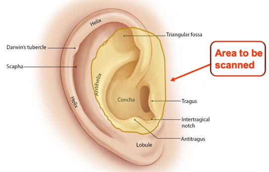
Otoscan - 3D Ear Scanning: The Future is Now!

Learn how 3D scanned ear images are used as a tool during hearing aid consultations to engage, educate, motivate, potentially improve outcomes, communicate value and differentiate your practice.

from #Audiology via ola Kala on Inoreader http://bit.ly/2D4WnCy
via IFTTT

A prospective analysis of motion and deformity at the shoulder level in surgically treated adolescent idiopathic scoliosis

Publication date: March 2019

Source: Gait & Posture, Volume 69

Author(s): R.M. Holewijn, M. de Kleuver, I. Kingma, N.L.W. Keijsers

Abstract
Background

Although posterior spinal correction and fusion surgery (PSF) of adolescent idiopathic scoliosis (AIS) limits counter rotation between thorax and pelvis, the physical function, and more specifically gait of these patients is only slightly affected after PSF. Possibly, shoulders-thorax counter-rotation increases to compensate for the loss in thorax-pelvis motion. This would subsequently result in a higher phase-difference and range of motion (ROM) between the shoulders and thorax.

Research questions

What is the effect of PSF on the phase difference and ROM between the shoulders and thorax? What is the effect of PSF on upper body deformity?

Methods

18 AIS patients underwent gait analysis at increasing walking speeds (0.45 to 2.22 m/s) before, and 3 and 12 months after PSF. The phase difference, ROM, and deformity between the shoulders, thorax, and pelvis were calculated.

Results

The shoulders- thorax phase difference was unaffected by surgery. At 3 months postoperatively the shoulders-thorax ROM was decreased (3.5° ± 0.2° versus 2.7° ± 0.2°, p=0.001). This recovered to preoperative values 12 months postoperatively (3.2° ± 0.2°, p=0.213). The shoulder-pelvis phase difference was decreased 3 months postoperatively (−98.9° ± 6.8° vs. −77.2° ± 7.2°, p=0.010), and recovered to pre-op values at the 12 months postoperative measurement (−89.6° ± 6.9°, p=0.290). Walking speed did not influence the effect of surgery on phase difference or ROM. The pre-operative shoulders-thorax asymmetry decreased from 3.4° ± 2.4° to 0.6° ± 3.1° (p<0.001). Shoulders-pelvis and thorax-pelvis asymmetry decreased from 10.0° ± 3.7° to 2.8° ± 4.3° (p<0.001) and from 6.5° ± 3.4° to 1.8° ± 3.2° (p=0.006) respectively.

Significance

No compensatory mechanisms could be identified in the relative motion between the shoulders and the thorax. Possibly, compensatory mechanisms are not required for normal gait after surgery. The asymmetry of the shoulders in the transversal plane improved without specific surgical strategies.


from #Audiology via ola Kala on Inoreader http://bit.ly/2UCHkHb