Τρίτη 12 Ιουλίου 2022

Understanding Etiologic Pathways Through Multiple Sequential Mediators: An Application in Perinatal Epidemiology

alexandrossfakianakis shared this article with you from Inoreader

SocialThumb.00001648.DC.jpeg

Background: Causal mediation analysis facilitates decomposing the total effect into a direct effect and an indirect effect that operates through an intermediate variable. Recent developments in causal mediation analysis have clarified the process of evaluating how – and to what extent – different pathways via multiple causally ordered mediators link the exposure to the outcome. Methods: Through an application of natural effect models for multiple mediators, we show how placental abruption might affect perinatal mortality using small-for-gestational-age (SGA) birth and preterm delivery as two sequential mediators. We describe methods to disentangle the total effect into the proportions mediated via each of the sequential mediators, when evaluating natural direct and natural indirect effects. Results: Under the assumption that SGA births causally precedes preterm delivery, an analysis of 16.7 million singleton pregnancies is consistent with the hypothesis that abruption exerts powerful effects on perinatal mortality (adjusted risk ratio=11.9; 95% confidence interval 11.6, 12.1). The proportions of the estimated total effect mediated through SGA birth and preterm delivery were 2% and 58%, respectively. The proportion unmediated via either SGA or preterm delivery was 41%. Conclusions: Through an application of causal mediation analysis with sequential mediators, we uncovered new insights into the pathways along which abruption impacts perinatal mortality. Copyright © 2022 Wolters Kluwer Health, Inc. All rights reserved.
View on Web

The Problem of Appetite Loss After Major Abdominal Surgery: A Systematic Review

alexandrossfakianakis shared this article with you from Inoreader
imageObjective: To systematically review the problem of appetite loss after major abdominal surgery. Summary of Background Data: Appetite loss is a common problem after major abdominal surgery. Understanding of etiology and treatment options is limited. Methods: We searched Medline, Cochrane Central Register of Controlled Trials, and Web of Science for studies describing postoperative appetite loss. Data were extracted to clarify definition, etiology, measurement, surgical influence, pharmacological, and nonpharmacological treatment. PROSPERO registration ID: CRD42021224489. Results: Out of 6144 articles, we included 165 studies, 121 of which were also analyzed quantitatively. A total of 19.8% were randomized, controlled trials (n = 24) and 80.2% were nonrandomized studies (n = 97). The studies included 20,506 patients undergoing the following surgeries: esophageal (n = 33 studies), gastric (n = 48), small bowel (n = 6), colon (n = 27), rectal (n = 20), hepatobiliary (n = 6), and pancreatic (n = 13). Appetite was mostly measured with the Quality of Life Questionnaire of the European Organization for Research and Treatment of Cancer (EORTC QLQ C30, n = 54). In a meta-analysis of 4 randomized controlled trials gum chewing reduced time to first hunger by 21.2 hours among patients who had bowel surgery. Other reported treatment options with positive effects on appetite but lower levels of evidence include, among others, intravenous ghrelin administration, the oral Japanese herbal medicine Rikkunshito, oral mosapride citrate, multidisciplin-ary-counseling, and watchin g cooking shows. No studies investigated the effect of well-known appetite stimulants such as cannabinoids, steroids, or megestrol acetate on surgical patients. Conclusions: Appetite loss after major abdominal surgery is common and associated with increased morbidity and reduced quality of life. Recent studies demonstrate the influence of reduced gastric volume and ghrelin secretion, and increased satiety hormone secretion. There are various treatment options available including level IA evidence for postoperative gum chewing. In the future, surgical trials should include the assessment of appetite loss as a relevant outcome measure.
View on Web

Δευτέρα 11 Ιουλίου 2022

Extramedullary focal fat - fluid level, a specific sign of osteomyelitis

Balaji Anvekar's Neuroradiology Cases

________________________________

Extramedullary focal fat - fluid level, a specific sign of osteomyelitis

Posted: 11 Jul 2022 09:37 PM PDT

 A 14 yo male with pain in calcaneum since 1months.


MRI foot for calcaneum with CT correlation shows:

Heterogeneous signal abnormality diffusely involving calcaneum with multiple low signal intensity foci diffusely scattered in calcaneum on T1-weighted images which are hyperintense on STIR. Rest of the intervening calcaneum medulla shows faint high signal on STIR.

No obvious density abnormality on CT. No obvious sclerotic or lytic lesion. No obvious cortical destruction or sclerosis.

There is a focal lentiform shaped parosteal collection measuring approximately 26 mm in height and 6 mm in thickness medially at 2 o'clock position and 4 mm in thickness laterally on plantar aspect at 7 o'clock position on axial section.

There is fat – fluid level within this collection, focal fat in the supernatant portion of this collection which is hyperintense on T1-weighted images with complete signal suppression on STIR, this portion follows classical fat density on CT.

There is an associated diffuse oedema involving muscles of plantar aspect of foot especially quadratus plantae muscle, medial as well as lateral teno synovitis.

This finding suggestive of extramedullary focal fat - fluid level which is a pathognomonic sign of acute to subacute osteomyelitis.

Findings were discussed with the referring physician before finalizing the report, who added that there is elevation of inflammatory markers in lab reports and the suspicion of osteomyelitis clinically as well, with a feedback of significant improvement clinically after IV antibiotics.

Take home note is during MSK MRI interpretation, a bone marrow signal abnormality with an associated focal periosteal extra medullary fat – fluid level, osteomyelitis should be in the list of your differential diagnosis.

References:

1. Extra-osseous fat fluid level: a specific sign for osteomyelitis. Kumar J, Bandhu S, Kumar A, Alam S. Skeletal Radiol. 2007 Jun;36 Suppl 1:S101-4. doi: 10.1007/s00256-006-0194-1. Epub 2006 Sep 19.

2. Intramedullary and extramedullary fat globules on magnetic resonance imaging as a diagnostic sign for osteomyelitis. Davies AM, Hughes DE, Grimer RJ. Eur Radiol. 2005 Oct;15(10):2194-9. doi: 10.1007/s00330-005-2771-4. Epub 2005 Apr 29.

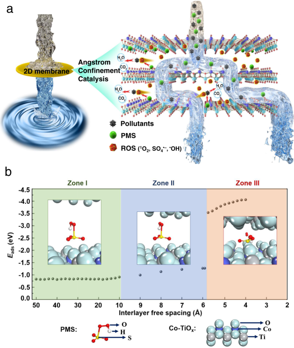
Angstrom-confined catalytic water purification within Co-TiOx laminar membrane nanochannels

alexandrossfakianakis shared this article with you from Inoreader

41467_2022_31807_Fig1_HTML.png

View on Web

Κυριακή 10 Ιουλίου 2022

Real‐life impact of tenofovir disoproxil fumarate and entecavir therapy on lipid profile, glucose and uric acid in chronic hepatitis B patients

alexandrossfakianakis shared this article with you from Inoreader

Abstract

Background & Aims

The impact of long-term nucleos(t)ide analogs treatment on host metabolism is a concern. Hence, we conducted this study to compare the effect of entecavir (ETV) and tenofovir disoproxil fumarate (TDF) on metabolic parameters among CHB patients.

Methods

In this real-life retrospective study, 2,030 CHB outpatients treated with ETV or TDF at Nanfang Hospital, China, were included. For treatment-naïve patients, pretreatment and semiannual metabolic parameters were collected. For treatment-experienced patients, metabolic parameters were collected at the first visit. Propensity score matching (PSM) was used to balance the effects of potential confounding factors.

Results

Among 122 treatment-naïve patients and 1908 treatment-experienced patients, ETV-treated patients were older with a higher percentage of metabolic syndrome. After PSM, the characteristics were comparable between the two groups. For treatment-naïve patients, fou r lipid parameters, including total cholesterol (TC), low-density lipoprotein (LDL), high-density lipoprotein and triglyceride levels showed a decreasing trend during the 42-month TDF treatment, while they remained relatively stable or increased during ETV treatment. At month 30, the levels of TC and LDL among TDF-treated patients were significantly lower than those among ETV-treated patients (TC: 4.7 vs. 3.9 mmol/L, p=0.004; LDL: 3.0 vs. 2.4 mmol/L, p=0.009). For treatment-experienced patients, we also observed lower levels of lipid parameters in patients with different durations of TDF treatment. The levels of glucose and uric acid were similar among ETV- and TDF-treated patients.

Conclusion

TDF has a lipid-lowering effect in CHB patients, which provides a basis for the selection of antiviral drugs for aging CHB patients.

This article is protected by copyright. All rights reserved.

View on Web

Longitudinal follow‐up of HPV16 sequence after cervical infection: low intra‐host variation and no correlation with clinical evolution

alexandrossfakianakis shared this article with you from Inoreader

ABSTRACT

Human papillomavirus (HPV) 16 exhibits different variants that may differ in their carcinogenic risk. In order to identify some high-risk variants, we sequenced and compared HPV16 whole genomes obtained from a longitudinal cohort of 34 HPV16-infected women who had either spontaneously cleared their infection (clearance group or "C"), or developed cervical high-grade lesions following a viral persistence (group persistence or "P"). Phylogenetic analysis of paired samples obtained at the beginning (C0 or P0) and at the end (C2 or P2) of the follow-up (median intervals between C0-C2 and between P0-P2 were 16 and 36.5 months, respectively) revealed a low genetic variability within the host compared to the genetic inter-host diversity. By comparing our HPV16 sequences to a reference sequence, we observed 301 different substitutions, more often transitions (60.9%) than transversions (39.1%), that occurred throughout the viral genome, but with a low frequen cy in E6 and E7 oncogenes (10 and 9 substitutions), suggesting a high conservation of these genes. Deletions and insertions were mostly observed in intergenic regions of the virus. The only significant substitution found between the subgroups C2 and P2 was observed in the L2 gene (L330F), with an unclear biological relevance. Our results suggest a low longitudinal intra-host evolution of HPV16 sequences and no correlation between genetic variations and clinical evolution.

This article is protected by copyright. All rights reserved.

View on Web

Influence of supplement administration of omega‐3 on the subcutaneous tissue response of endodontic sealers in Wistar Rats

alexandrossfakianakis shared this article with you from Inoreader

Abstract

Aim

Natural substances such as omega-3 have been used in the medical field due to their numerous properties and, in particular, modulating effect on the systemic and local inflammatory processes. Thus, this study evaluated the influence of omega-3 supplementation on the subcutaneous tissue response of endodontic sealers in Wistar Rats.

Methodology

Polyethylene tubes were implanted in the subcutaneous tissue of 48 animals (one empty for control and three filled with Sealapex, AH Plus or Endofill). The animals were treated with omega-3 (TO) or water (TW). Treatments started 15 days before implantation until euthanasia. After 5, 15 and 30 days (n=8), animals were euthanized and polyethylene tubes and surrounding tissue were removed and processed for histological analysis. The inflammatory reaction was analysed by Haematoxylin and Eosin stain and immunolabeling for IL-6 and TNF-α. The collagen maturity was analysed by picrosirius red stain and calcium deposition by von Kossa stain and polarized light. Results were statistically analysed (p<0.05).

Results

Among TW sealers groups, Endofill evoked a more intense inflammatory infiltrate compared with AH Plus and control in the 30-day period (p=0.009). However, in TO sealers groups, there was no difference among the sealers and control in all periods (p>0.05). Comparing each sealer as a function of the supplementation with water or omega-3, there are differences for Endofill (p=0.001) and Sealapex (p=0.005) in the 30-day period, presenting lower inflammatory infiltrate in the animals treated with omega 3. A higher percentage of immature fibres was observed at 15 and 30 days in the TO group, compared with TW group (p<0.05). The deposition of calcium particles was observed only by Sealapex in all periods, despite the supplementation procedure.

Conclusions

Omega-3 supplementation influence the tissue reactions of endodontic sealers, modulating inflammation, the immunolabeling of IL-6 and TNF-α, the repair process and it does not interfere with calcium deposition.

View on Web