Δευτέρα 26 Απριλίου 2021

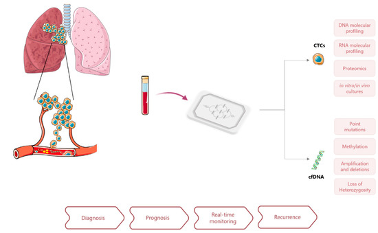
Cancers, Vol. 13, Pages 2101: Emerging Lab-on-a-Chip Approaches for Liquid Biopsy in Lung Cancer: Status in CTCs and ctDNA Research and Clinical Validation

xloma.fota13 shared this article with you from Inoreader
Via Cancers

cancers-13-02101-g001-550.jpg

Cancers, Vol. 13, Pages 2101: Emerging Lab-on-a-Chip Approaches for Liquid Biopsy in Lung Cancer: Status in CTCs and ctDNA Research and Clinical Validation

Cancers doi: 10.3390/cancers13092101

Authors: Ângela Carvalho Gabriela Ferreira Duarte Seixas Catarina Guimarães-Teixeira Rui Henrique Fernando J. Monteiro Carmen Jerónimo

Despite the intensive efforts dedicated to cancer diagnosis and treatment, lung cancer (LCa) remains the leading cause of cancer-related mortality, worldwide. The poor survival rate among lung cancer patients commonly results from diagnosis at late-stage, limitations in characterizing tumor heterogeneity and the lack of non-invasive tools for detection of residual disease and early recurrence. Henceforth, research on liquid biopsies has been increasingly devoted to overcoming these major limitations and improving management of LCa patients. Liquid biopsy is an emerging field that has evolved significantly in recent years due its minimally invasive nature and potential to assess various disease biomarkers. Several strategies for characterization of circulating tumor cells (CTCs) and circulating tumor DNA (ctDNA) have been developed. With the aim of standardizing diagnostic and follow-up practices, microfluidic devices have been introduced to improve biomarkers isolation efficiency and specificity. Nonetheless, implementation of lab-on-a-chip platforms in clinical practice may face some challenges, considering its recent application to liquid biopsies. In this review, recent advances and strategies for the use of liquid biopsies in LCa management are discussed, focusing on high-throughput microfluidic devices applied for CTCs and ctDNA isolation and detection, current clinical validation studies and potential clinical utility.

View on the web

Correlation of Voice Hoarseness and Vocal Cord Palsy: A Prospective Assessment of Recurrent Laryngeal Nerve Injury Following Anterior Cervical Discectomy and Fusion

xloma.fota13 shared this article with you from Inoreader

Int J Spine Surg. 2021 Feb;15(1):12-17. doi: 10.14444/8001. Epub 2021 Feb 12.

ABSTRACT

BACKGROUND: Injury to the recurrent laryngeal nerve (RLN) has been implicated as a common complication following anterior cervical discectomy and fusion (ACDF) surgery. The purpose of this study is to determine the true incidence of voice hoarseness and RLN palsy following ACDF surgery, to determine the reliability of symptoms in the diagnosis of RLN injury, and to evaluate factors related to the development of these symptoms.

METHODS: All patients undergoing elective (primary or secondary) ACDF surgery at a single institution consented to and enrolled in the present study. All approaches were through the left side. Enrolled patients received both preoperative and postoperative (within 1 month following surgery) laryngoscopy by a fellowship-trained ENT physician for evaluation of RLN function. Patients also responded as to whether they were experienci ng postoperative symptoms of dysphagia, aspiration, and voice changes.

RESULTS: In total, 108 patients were included in this study. Mean age of the population was 59.2 ± 10.7 years and mean body mass index was 31.2 ± 7.1 kg/m2. Three patients had previously undergone a thyroidectomy, whereas 20 patients had undergone a previous ACDF. Average intubation time for ACDF surgery was 121.6 ± 38.5 minutes. After surgery and excluding patients who were experiencing preoperative symptoms, 19 patients (20.4%) complained of dysphagia, 2 patients (1.9%) complained of aspiration symptoms, and 5 patients (4.6%) complained of voice hoarseness. There was no incidence of vocal cord palsy from postoperative laryngoscopy. From multivariate analysis, endotracheal cuff pressure after retractor placement was correlated to postoperative voice hoarseness, dysphagia, and aspiration symptoms.

CONCLUSIONS: From the results of this prospective study, the RLN remained functional even a month after surgery despite several cases of postoperative dysphagia, aspiration, and voice changes. Endotracheal cuff pressure, number of vertebral levels, body mass index, and intubation time were important variables related to postoperative symptoms.

CLINICAL RELEVANCE: Voice hoarseness does not necessarily indicate recurrent laryngeal nerve injury after ACDF but may be caused by compressive forces on laryngeal tissue during retraction or intubation. Laryngoscopy should be performed in cases with high clinical suspicion.

LEVEL OF EVIDENCE: 2.

PMID:33900952 | DOI:10.14444/8001

View on the web

Web-Based Educational Intervention for Patients With Uninvestigated Dyspepsia Referred for Upper Gastrointestinal Tract Endoscopy: A Randomized Clinical Trial

xloma.fota13 shared this article with you from Inoreader

JAMA Intern Med. 2021 Apr 26. doi: 10.1001/jamainternmed.2021.1408. Online ahead of print.

ABSTRACT

IMPORTANCE: Diagnostic yield of upper gastrointestinal (GI) tract endoscopy for uninvestigated dyspepsia is low, and its clinical implications are limited. There is an unmet need for better strategies to reduce the volume of upper GI tract endoscopic procedures for dyspepsia.

OBJECTIVE: To study the effectiveness of a web-based educational intervention as a tool to reduce upper GI tract endoscopy in uninvestigated dyspepsia.

DESIGN, SETTING, AND PARTICIPANTS: This open-label, multicenter, randomized clinical trial enrolled participants between November 1, 2017, and March 31, 2019, with follow-up 52 weeks after randomization, at 4 teaching hospitals in the Netherlands. Participants included patients with uninvestigated dyspeptic symptoms who were referred for upper GI tract endoscopy by their general health care clinician without pri or consultation of a gastroenterologist. A total of 119 patients, aged 18 to 69 years, were included. Patients were excluded if any of the following red flag symptoms were present: (indirect) signs of upper GI tract hemorrhage (hematemesis, melena, hematochezia, or anemia), unintentional weight loss of 5% or higher of normal body weight during a period of 6 to 12 months, persistent vomiting, dysphagia, or jaundice.

INTERVENTIONS: Patients were randomly assigned (1:1) to education (intervention) or upper GI tract endoscopy (control). Education consisted of a self-managed web-based educational intervention, containing information on gastric function, dyspepsia, and upper GI tract endoscopy.

MAIN OUTCOMES AND MEASURES: Difference in the proportion of upper GI tract endoscopy procedures between those who received access to the web-based educational intervention and those who did not at 12 weeks and 52 weeks after randomization, analyzed in the intention-to-treat population. Secondary outcomes included quality of life (Nepean Dyspepsia Index) and symptom severity (Patient Assessment of Gastrointestinal Disorders Symptom Severity Index) measured at baseline and 12 weeks.

RESULTS: Of 119 patients included (median age, 48 years [interquartile range, 37-56 years]; 48 men [40%]), 62 were randomized to web-based education (intervention) and 57 to upper GI tract endoscopy (control). Significantly fewer patients compared with controls underwent upper GI tract endoscopy after using the web-based educational intervention: 24 (39%) vs 47 (82%) (relative risk, 0.46; 95% CI, 0.33-0.64; P < .001). Symptom severity and quality of life improved equivalently in both groups. One additional patient in the intervention group required upper GI tract endoscopy during follow-up.

CONCLUSIONS AND RELEVANCE: Findings of this study indicate that web-based patient education is an effective tool to decrease the need for upper GI tract endoscopy in uninvestigated dysp epsia.

TRIAL REGISTRATION: ClinicalTrials.gov Identifier: NCT03205319.

PMID:33900373 | DOI:10.1001/jamainternmed.2021.1408

View on the web

Κυριακή 25 Απριλίου 2021

Combined immune checkpoint inhibitors of CTLA4 and PD-1 for hepatic melanoma of unknown primary origin: A case report

paythelady.612 shared this article with you from Inoreader

World J Clin Cases. 2021 Apr 16;9(11):2641-2648. doi: 10.12998/wjcc.v9.i11.2641.

ABSTRACT

BACKGROUND: Melanoma is uncommonly found in lymph nodes, subcutaneous tissue, or visceral organs without a primary lesion, where it is identified as metastatic melanoma with unknown primary (MUP). Hepatic MUP is extremely rare and has a poor prognosis. There is limited information on its pathogenesis, clinical and imaging features, and pathological findings. There are no guidelines for the use of immune checkpoint inhibitors (ICIs) in hepatic MUP, and the treatment outcome has rarely been reported.

CASE SUMMARY: A 42-year-old woman presented to our hospital with hepatic tumors found incidentally during a routine check-up. Contrast-enhanced abdominal com-puterized tomography showed multiple mass lesions in the liver. Pathological results revealed melanoma, which was confirmed by immunohistochemical staining for HMB-45(+), Melan-A(+), S-100(+) , and SOX10(+). There was no evidence of primary cutaneous, ocular, gastrointestinal, or anal lesion on a comprehensive examination. The patient was diagnosed with hepatic MUP. She received combined antibodies against cytotoxic T-lymphocyte-associated antigen 4 (CTLA-4, ipilimumab) and programmed death protein-1 (PD-1, nivolumab). She died of hepatic failure 9 mo after hepatic MUP was diagnosed. This the first case of hepatic MUP treated with combined ipilimumab and nivolumab, who showed better outcome than previous cases.

CONCLUSION: Combined ICIs of PD-1 and CTLA-4 may be considered as the first-line therapy for patients with hepatic MUP.

PMID:33889631 | PMC:PMC8040165 | DOI:10.12998/wjcc.v9.i11.2641

View on the web

Cerebellar artery infarction with sudden hearing loss and vertigo as initial symptoms: A case report

paythelady.612 shared this article with you from Inoreader

World J Clin Cases. 2021 Apr 16;9(11):2519-2523. doi: 10.12998/wjcc.v9.i11.2519.

ABSTRACT

BACKGROUND: Sudden hearing loss (SHL) is associated with serious systematic conditions such as neoplasms, vascular events, autoimmune diseases, infections, and iatrogenic injury. Some authors report that SHL can be an early warning sign of impending vertebrobasilar ischemic stroke. It is important to distinguish stroke from benign disease.

CASE SUMMARY: A 48-year-old male patient presented with SHL and vertigo as first symptoms. Diffusion-weighted imaging revealed high signal intensity in the left posterior inferior cerebellar artery territory of the cerebellar hemisphere and high signal intensity in the right pons and bridge cerebellar arm, confirming that the patient had cerebral infarction. Treatment with antiplatelet drugs, steroid anti-inflammatory drugs, and neurotrophic nerve therapy promoted blood circulation and removed blood stasis , and the symptoms of the patient were significantly improved.

CONCLUSION: SHL and vertigo could be the initial symptoms of vertebrobasilar ischemic stroke.

PMID:33889616 | PMC:PMC8040184 | DOI:10.12998/wjcc.v9.i11.2519

View on the web

Cholangiojejunostomy for multiple biliary ducts in living donor liver transplantation: A case report

paythelady.612 shared this article with you from Inoreader

World J Clin Cases. 2021 Apr 16;9(11):2649-2654. doi: 10.12998/wjcc.v9.i11.2649.

ABSTRACT

BACKGROUND: Laparoscopic living donor hepatectomy (LLDH) has been successfully carried out in several transplant centers. Biliary reconstruction is key in living donor liver transplantation (LDLT). Reliable biliary reconstruction can effectively prevent postoperative biliary stricture and leakage. Although preoperative magnetic resonance cholangiopancreatography and intraoperative indocyanine green cholangiography have been shown to be helpful in determining optimal division points, biliary variability and limitations associated with LLDH, multiple biliary tracts are often encountered during surgery, which inhibits biliary reconstruction. A reliable cholangiojejunostomy for multiple biliary ducts has been utilized in LDLT. This procedure provides a reference for multiple biliary reconstructions after LLDH.

CASE SUMMARY: A 2-year-old girl dia gnosed with ornithine transcarbamylase deficiency required liver transplantation. Due to the scarcity of deceased donors, she was put on the waiting list for LDLT. Her father was a suitable donor; however, after a rigorous evaluation, preoperative magnetic resonance cholangiopancreatography examination of the donor indicated the possibility of multivessel variation in the biliary tract. Therefore, a laparoscopic left lateral section was performed on the donor, which met the estimated graft-to-recipient weight ratio. Under intraoperative indocyanine green cholangiography, 4 biliary tracts were confirmed in the graft. It was difficult to reform the intrahepatic bile ducts due to their openings of more than 5 mm. A reliable cholangiojejunostomy was, therefore, utilized: Suture of the jejunum to the adjacent liver was performed around the bile duct openings with 6/0 absorbable sutures. At the last follow-up (1 year after surgery), the patient was complication-free.

CONCLUSION: Int rahepatic cholangiojejunostomy is reliable for multiple biliary ducts after LLDH in LDLT.

PMID:33889632 | PMC:PMC8040162 | DOI:10.12998/wjcc.v9.i11.2649

View on the web

Extraosseous spinal epidural plasmocytoma associated with multiple myeloma: Two case reports

paythelady.612 shared this article with you from Inoreader

World J Clin Cases. 2021 Apr 16;9(11):2555-2561. doi: 10.12998/wjcc.v9.i11.2555.

ABSTRACT

BACKGROUND: Multiple myeloma is a malignant hematological disease characterized by proliferation of monoclonal plasma cells mainly in the bone marrow. Extraosseous epidural plasmacytoma associated with myeloma arises from lymphoid tissue in the epidural space without focal vertebral involvement, and is rare.

CASE SUMMARY: A 52-year-old woman was diagnosed with kappa subtype nonsecretory multiple myeloma and presented with bilateral arm weakness 11 mo after completing multiple courses of chemotherapy. Spinal magnetic resonance imaging (MRI) showed a posterior C7-T3 epidural mass with spinal cord compression. After five courses of chemotherapy, follow-up MRI showed resolution of cord compression. A 54-year-old man presented with paraplegia 15 mo after a diagnosis of IgD kappa subtype multiple myeloma and completing multiple courses of chemothe rapy. He underwent Th11 and L1 laminectomies for tumor resection because MRI showed an epidural mass causing cord compression. His-topathologic examination was consistent with IgD multiple myeloma. The patients have currently survived for 33 mo and 19 mo, respectively.

CONCLUSION: Isolated extraosseous epidural plasmacytoma associated with multiple myeloma without bony involvement is difficult to diagnose by imaging. Definitive diagnosis requires pathological and immunohistochemical examination.

PMID:33889620 | PMC:PMC8040174 | DOI:10.12998/wjcc.v9.i11.2555

View on the web