Δευτέρα 28 Φεβρουαρίου 2022

Quality of Life and Functionality of Head and Neck Cancer Patients Are Diminished As a Function of Sarcopenia and Obesity

xlomafota13 shared this article with you from Inoreader

pubmed-meta-image.png

Ear Nose Throat J. 2022 Feb 28:1455613221076791. doi: 10.1177/01455613221076791. Online ahead of print.

ABSTRACT

BACKGROUND: Health-related quality of life (QoL) is a measure that allows us to know the patient's perception of well-being and how it is affected by their disease and treatments. In cancer patients, sarcopenia has been associated with low scores on various instruments used to assess the QoL; however, little information is available on the effects of sarcopenia an d sarcopenic obesity on the QoL of patients with head and neck cancer (H&NC).

METHODS: In this cross-sectional study with 71 H&NC patients aged between 40 and 80 years, we describe the scores on the instruments EORTC QLQ C-30 and EORTC QLQ-H&N35 according to the sarcopenia phenotype (NSG, nonsarcopenic group; SG, sarcopenic group; and SOG, sarcopenic obesity group), hand-grip strength, gait speed, total lymphocyte count, albumin, cholesterol and C-reactive protein, and the relationships between these variables.

RESULTS: The prevalence of sarcopenia and sarcopenic obesity was 48% and 28%, respectively. The QoL analysis showed that NSG had higher scores on the physical functioning scale [NSG 93 (83-100); SG 73 (52-88); SOG 83 (53-93), P = .009] and lower scores on the fatigue scale [NSG 11 (0-22); S 39 (30-67); SOG 44 (14-56); P = .004]. The NSG had a higher hand-grip strength (31.1 kg) than SG (24.1 kg, P = .007) and SOG (26.3 kg, P = .001), and a lower C-reac tive protein. The SG and SOG showed no differences between them.

CONCLUSIONS: Patients with sarcopenia or sarcopenic obesity have lower physical performance and a higher level of fatigue than nonsarcopenic patients. This loss of function can maintain or worsen sarcopenia due to the patient's self-restraint in physical exertion that encourages an increase in muscle tissue.

PMID:35226551 | DOI:10.1177/01455613221076791

View on the web

Κυριακή 27 Φεβρουαρίου 2022

Can cystic lesions of the jaws be considered as the cause of mandibular asymmetry?

xlomafota13 shared this article with you from Inoreader

Med Oral Patol Oral Cir Bucal. 2022 Mar 1;27(2):e159-e163. doi: 10.4317/medoral.25134.

ABSTRACT

BACKGROUND: The aim of this study is to investigate the presence of condylar and ramal asymmetry in patients with a cyst larger than 10 mm in the maxilla or mandible.

MATERIAL AND METHODS: Condylar and ramal asymmetry index measurements of 47 patients (mean age: 28.85 ± 15.348) in the study group and 40 patients in the control group (mean age: 33.73 ± 13.095) were performed using panoramic radiographs. The study group consists of patients with cysts larger than 10 mm in diameter in the maxilla or mandible. The control group consisted of patients with no radiolucent lesions and no history of trauma. The possible statistical difference between the groups was evaluated by the Mann-Whitney U test.

RESULTS: No statistically significant difference was observed in asymmetry indices according to gender and the jaw (maxilla or mandible) in which the cyst was located. However, it was determined that CAI and RAI values were statistically significantly different between the study and control groups (p = 0.047 and p = 0.016, respectively).

CONCLUSIONS: The presence of intraosseous cysts larger than 10 mm in the jaws was found to be associated with condylar and ramal asymmetry.

PMID:35218644 | DOI:10.4317/medoral.25134

View on the web

Πέμπτη 24 Φεβρουαρίου 2022

Traumatic pseudoaneurysm of posterior superior alveolar artery with epistaxis as a main symptom: a case report

xlomafota13 shared this article with you from Inoreader

Zhonghua Er Bi Yan Hou Tou Jing Wai Ke Za Zhi. 2022 Feb 7;57(2):201-203. doi: 10.3760/cma.j.cn115330-20210407-00185.

NO ABSTRACT

PMID:35196765 | DOI:10.3760/cma.j.cn115330-20210407-00185

View on the web

Phase II study of apatinib combined with temozolomide in patients with advanced melanoma after failure of immunotherapy

xlomafota13 shared this article with you from Inoreader

SocialThumb.00008390.DC.jpeg

Objective Treatment for advanced melanoma after progression on immunotherapy is limited. This phase II trial (NCT03422445) was conducted to evaluate the efficacy and safety of apatinib plus temozolomide in patients with advanced melanoma after failure of immunotherapy. Methods Patients with unresectable stage III or stage IV melanoma after progression on immunotherapy were treated with temozolomide 300 mg on days 1–5 and apatinib 500 mg daily every 28-day cycle until disease progression or intolerable toxicities. Besides immunotherapy, prior chemotherapy, targeted therapy, and clinical trials were allowed. The primary endpoint was progression-free survival. Secondary endpoints were objective response rate, disease control rate, overall survival, and safety. Results Of 29 patients, 28 (96.6%) had metastatic diseases, and the predominant subtypes were mucosal [12 (41.4%)] and acral melanoma [eight (27.6%)]. Five (17.2%) patients showed BRAF, CKIT, or NRAS mutation. Five achieved confirmed partial response, with an objective response rate of 17.2%. The disease control rate was 82.8%. The median progression-free survival was 5.0 months [95% confidence interval (CI): 4.7–5.3], and the median overall survival was 10.1 months (95% CI: 5.1–15.0). Grade 3–4 treatment-related adverse events included proteinuria [four (13.8%)], thrombocytopenia [two (6.9%)], hypertension [one (3.4%)], and hyperbilirubinemia [one (3.4%)]. No treatment-related death occurred. Conclusion Apatinib plus temozolomide demonstrated promising efficacy and manageable safety profile in patients with advanced melanoma after progression on immunotherapy. * Li Zhou and Yue Yang contributed equally to the writing of this article and are the co-first authors. Supplemental Digital Content is available for this article. Direct URL citations appear in the printed text and are provided in the HTML and PDF versions of this article on the journal's website, www.melanomaresearch.com. Received 11 November 2021 Accepted 25 January 2022 Correspondence to Chuanliang Cui, Department of Renal Cancer and Melanoma, Peking University Cancer Hospital and Institute, Beijing, China, Tel: +8610 88196317 -mail: 1008ccl@163.com Copyright © 2022 Wolters Kluwer Health, Inc. All rights reserved.
View on the web

Τετάρτη 23 Φεβρουαρίου 2022

Similarities and differences of estrogen in the regulation of temporomandibular joint osteoarthritis and knee osteoarthritis

xlomafota13 shared this article with you from Inoreader

Histol Histopathol. 2022 Feb 23:18442. doi: 10.14670/HH-18-442. Online ahead of print.

ABSTRACT

BACKGROUND: Temporomandibular joint osteoarthritis (TMJOA) and knee osteoarthritis (knee OA) are two kinds of common osteoarthritis (OA) that are characterized by chronic degeneration of soft and hard tissues around joints. Their gender and age differences suggest that there are similarities and differences between the pathogenic mechanisms of TMJOA and knee OA.

OBJECTIVE: To review recent studies on the effect of estrogen on TMJOA and knee OA, and summarize their possible pathogenesis and molecular mechanisms.

SOURCES: Articles up to present reporting the relationship of estrogen and TMJOA or knee OA are included. An extensive electronic search was conducted of databases including PubMed, Web of science core collection.

CONCLUSION: According to epidemiological investigations, TMJOA primarily happens to females of puberty a nd childbearing age, while knee OA mainly affects postmenopausal women. Epidemiological investigation and experimental research suggest that estrogen may have a different effect on TMJ and on knee. Though estrogen regulates TMJOA and knee OA via estrogen-related receptors (ERR), their pathogenesis and pathway of estrogen regulation are different. To find out the accurate regulation of estrogen on TMJOA and knee OA, specific pathways and molecular mechanisms still need further exploration.

PMID:35194774 | DOI:10.14670/HH-18-442

View on the web

Hyperintense areas in the cisternal segments of the cranial nerves: a magnetic resonance imaging study

xlomafota13 shared this article with you from Inoreader

pubmed-meta-image.png

Surg Radiol Anat. 2022 Feb 23. doi: 10.1007/s00276-022-02902-1. Online ahead of print.

ABSTRACT

PURPOSE: The study aimed to explore hyperintense areas in the cisternal segments of the cranial nerves using magnetic resonance imaging (MRI).

METHODS: Seventy outpatients underwent thin-sliced, coronal constructive interference steady-state (CISS) sequence and sagittal T2-weighted MRI following conventional MRI examination.

RESULTS: With the coronal CISS sequence, hype rintense areas were located in the central parts of the olfactory bulbs in 65.7% of patients. For the intracranial optic nerve and optic chiasm, hyperintense areas were detected in 98.6% of the CISS sequences and 100% of the T2-weighted images. In the optic tract, hyperintense areas were detected in 51.4% of cases. In 35% of the patients who underwent the CISS sequence, the intracranial optic nerves were considerably compressed by the internal carotid and anterior cerebral arteries, with hyperintense areas similar to those in patients without vascular compression. Hyperintense areas of the cisternal segments of the oculomotor nerve and trigeminal root were identified in 52.9% and 87.1% of the patients, respectively.

CONCLUSIONS: The hyperintense areas found within the cisternal segments of the cranial nerves delineated on the coronal CISS sequence and sagittal T2-weighted imaging may indicate the intracranial part of the glymphatic pathway through the cranial nerves. The crani al nerves may function as part of the glymphatic pathway.

PMID:35195771 | DOI:10.1007/s00276-022-02902-1

View on the web

Τρίτη 22 Φεβρουαρίου 2022

Selumetinib Plus Adjuvant Radioactive Iodine in Patients With High-Risk Differentiated Thyroid Cancer: A Phase III, Randomized, Placebo-Controlled Trial (ASTRA)

xlomafota13 shared this article with you from Inoreader

pubmed-meta-image.png

J Clin Oncol. 2022 Feb 22:JCO2100714. doi: 10.1200/JCO.21.00714. Online ahead of print.

ABSTRACT

PURPOSE: Selumetinib can increase radioactive iodine (RAI) avidity in RAI-refractory tumors. We investigated whether selumetinib plus adjuvant RAI improves complete remission (CR) rates in patients with differentiated thyroid cancer (DTC) at high risk of primary treatment failure versus RAI alone.

METHODS: ASTRA (NCT01843062) is an international, phase III, randomized, placebo-con trolled, double-blind trial. Patients with DTC at high risk of primary treatment failure (primary tumor > 4 cm; gross extrathyroidal extension outside the thyroid gland [T4 disease]; or N1a/N1b disease with ≥ 1 metastatic lymph node(s) ≥ 1 cm or ≥ 5 lymph nodes [any size]) were randomly assigned 2:1 to selumetinib 75 mg orally twice daily or placebo for approximately 5 weeks (no stratification). On treatment days 29-31, recombinant human thyroid-stimulating hormone (0.9 mg)-stimulated RAI (131I; 100 mCi/3.7 GBq) was administered, followed by 5 days of selumetinib/placebo. The primary end point (CR rate 18 months after RAI) was assessed in the intention-to-treat population.

RESULTS: Four hundred patients were enrolled (August 27, 2013-March 23, 2016) and 233 randomly assigned (selumetinib, n = 155 [67%]; placebo, n = 78 [33%]). No statistically significant difference in CR rate 18 months after RAI was observed (selumetinib n = 62 [40%]; placebo n = 30 [38%]; odds ratio 1.07 [95% CI, 0.61 to 1.87]; P = .8205). Treatment-related grade ≥ 3 adverse events were reported in 25/154 patients (16%) with selumetinib and none with placebo. The most common adverse event with selumetinib was dermatitis acneiform (n = 11 [7%]). No treatment-related deaths were reported.

CONCLUSION: Postoperative pathologic risk stratification identified patients with DTC at high risk of primary treatment failure, although the addition of selumetinib to adjuvant RAI failed to improve the CR rate for these patients. Future strategies should focus on tumor genotype-tailored drug selection and maintaining drug dosing to optimize RAI efficacy.

PMID:35192411 | DOI:10.1200/JCO.21.00714

View on the web

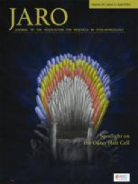
Cochlear Fluid Spaces and Structures of the Gerbil High-Frequency Region Measured Using Optical Coherence Tomography (OCT)

xlomafota13 shared this article with you from Inoreader

10162.jpg

Abstract

Since it has been difficult to directly observe the morphology of the living cochlea, our ability to infer the mechanical functioning of the living ear has been limited. Nearly all our knowledge about cochlear morphology comes from postmortem tissue that was fixed and processed using procedures that possibly distort the structures and fluid spaces of the organ of Corti. In this study, optical coherence tomography was employed to obtain volumetric images of the high-frequency hook region of the gerbil cochlea, as viewed through the round window, with far better resolution capability than had been possible before. The anatomical structures and fluid spaces of the organ of Corti were segmented and quantified in vivo and over a 90-min postmortem period. We find that the arcuate-zone and pectinate-zone widths change very little postmortem. The volume of the scala tympani between the round-window membrane and basilar membrane and the volume of the inner spiral sulcus d ecrease in the first 60-min postmortem. While textbook drawings of the mammalian organ of Corti and cortilymph prominently depict the tunnel of Corti, the outer tunnel is typically missing. This is likely because textbook drawings are typically made from images obtained by histological methods. Here, we show that the outer tunnel is nearly twice as big as the tunnel of Corti or the space of Nuel. This larger outer tunnel fluid space could have a substantial, little-appreciated effect on cochlear micromechanics. We speculate that the outer tunnel forms a resonant structure that may affect reticular-lamina motion.

View on the web

Δευτέρα 21 Φεβρουαρίου 2022

Diagnosis of mucormycosis using an intercalating dye-based quantitative PCR

xlomafota13 shared this article with you from Inoreader

Med Mycol. 2022 Feb 21:myac015. doi: 10.1093/mmy/myac015. Online ahead of print.

ABSTRACT

PCR-based methods applied to various body fluids emerged in recent years as a promising approach for the diagnosis of mucormycosis. In this study, we set up and assess the value of a qPCR to detect a wide variety of Mucorales species in a single tube. A pair of degenerated primers targeting the rDNA operon was used in a qPCR utilizing an intercalating fluorescent dye. Analytical assessment, using a wide variety of both Mucorales strains (8 genera, 11 species) and non-Mucorales strains (9 genera, 14 species), showed 100% sensitivity and specificity rates with a limit of detection at 3 rDNA copy/ qPCR reaction. Subsequently, 364 clinical specimens from 166 at-risk patients were prospectively tested with the assay. All the seven patients classified as proven/probable mucormycosis using the EORTC-MSG criteria had a positive qPCR as well as a patient with a p roven uncharacterized invasive mould infection. In addition, 3 out of 7 patients with possible mould invasive infections had at least one positive qPCR test. Sensitivity was calculated between 73.33% to 100% and specificity between 98.10% to 100%. The qPCR method proposed showed excellent performances and would be an important adjunctive tool for the difficult diagnosis of mucormycosis diagnosis.

PMID:35188208 | DOI:10.1093/mmy/myac015

View on the web

Cricotracheostomy in a patient with severe kyphosis: A case report

xlomafota13 shared this article with you from Inoreader

pubmed-meta-image.png

Ear Nose Throat J. 2022 Feb 21:1455613221077596. doi: 10.1177/01455613221077596. Online ahead of print.

ABSTRACT

Cricotracheostomy is a useful surgical procedure for opening the airway in cases where conventional tracheotomy is difficult. This is a safe and easy technique involving removal of the anterior portion of the cricoid cartilage. Herein, we report the case of a patient with difficulty in neck extension due to severe kyphosis, who underwent cricotracheostomy. A 65-ye ar-old woman developed dyspnea and was transported to our hospital. SpO2 was 60% (room air) and endotracheal intubation was performed. She was managed on ventilator. However, weaning off the ventilator was difficult, and endotracheal intubation was prolonged. Subsequently, the patient was referred to us for a tracheostomy. The patient was unable to maintain the supine position due to severe kyphosis, and computed tomography revealed that the brachiocephalic artery was running just below the thyroid gland. Therefore, conventional tracheostomy was difficult to perform, and we performed cricotracheostomy instead. After the cricotracheostomy, there were no tracheal problems, such as tracheal stenosis and stomal hemorrhage. Cricotracheostomy can be performed in cases where multiple anatomical difficulties may be faced in the tracheostomy site, as in this case. Furthermore, cricotracheostomy is beneficial for long-term airway management because it causes fewer complications com pared to conventional tracheotomy.

PMID:35188407 | DOI:10.1177/01455613221077596

View on the web

Κυριακή 20 Φεβρουαρίου 2022

Features of Odontogenic Sinusitis Associated With Dental Implants

xlomafota13 shared this article with you from Inoreader

Objectives

With the increase in dental implants for tooth loss, odontogenic sinusitis following maxillary dental implants is frequently encountered in otorhinolaryngology practice. The authors aimed to reveal the association between implant extrusion into maxillary sinus, along with implant-related complications in patients diagnosed with implant-related odontogenic sinusitis (IR-ODS).

Study Design

Case–control study.

Methods

This study enrolled 60 patients who received functional endoscopic sinus surgery due to IR-ODS. The preoperative sinus computed tomography was retrospectively reviewed. Among the 120 maxillary sinuses of the 60 patients, 68 sides were diagnosed with IR-ODS sides, whereas 27 sides showed no clinical or radiological evidence of this condition after the implant insertion and were defined as the control sides. Statistical analysis between these two groups was conducted, in addition to odds ratio (OR) calculations for associations with IR-ODS.

Results

The mean age of the IR-ODS subjects was 59.5 ± 19.1, with a male to female ratio of 32/28 (53.3%/46.7%). Implants extruding by more than 4 mm into the maxillary sinus, peri-implantitis, bone graft disruption–extrusion were associated with a significantly higher incidence in the IR-ODS (p = 0.035, p = 0.003, p = 0.011, respectively). The IR-ODS sides showed an adjusted-OR (95% confidence interval) of 27.4 (2.7–276.5) for extrusion length >4 mm, 11.8 (3.0–46.5) for peri-implantitis, and 34.1 (3.3–347.8) for bone graft disruption (p = 0.005, p < 0.001, and p = 0.003, respectively).

Conclusion

Maxillary dental implants extruding more than 4 mm into the maxillary sinus, peri-implantitis, and disrupted–extruded bone grafts show significant association with IR-ODS.

Level of Evidence

4 Laryngoscope, 2022

View on the web